February 10, 2025
New imaging technology at IU Health Saxony enhances safety for heart patients
IU Health Fishers
By Emma Avila, epackard1@iuhealth.org, writer for IU Health's Metro Region
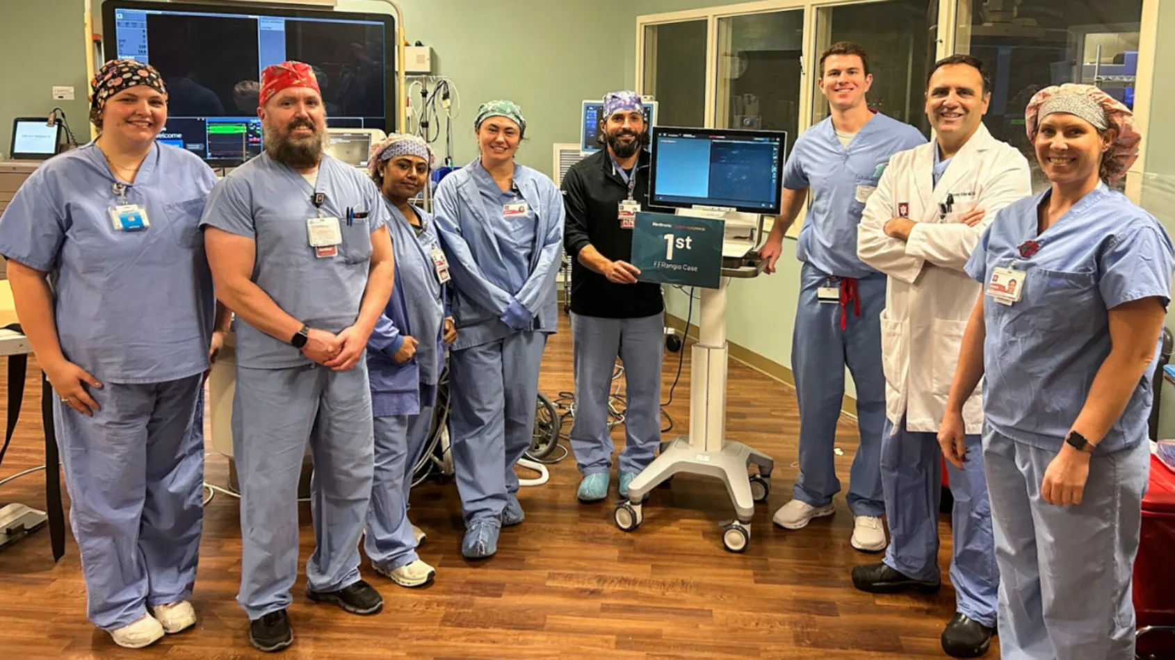
The Cath Lab team at IU Health Saxony Hospital is using new technology for angiograms, enhancing patient safety.
Dr. Ali Farooq Iqtidar, cardiologist, and the Cath Lab team at IU Health Saxony Hospital are leading the way in improving patient safety and outcomes with a new, less invasive imaging technology for heart patients. The process uses advanced computer learning to provide better, safer care for patients needing angiograms.
The
IU Health Saxony Cardiology team is the first in Hamilton County to adopt this
less invasive approach to angiogram analysis.
An angiogram is a type of X-ray that helps doctors see how blood is flowing, and it's crucial for spotting problems like blockages, aneurysms (blood vessel bulges) and blood clots. Historically, when patients have moderate blockages in the blood vessels of their heart, doctors insert a wire into the blood vessel to help determine the significance of these blockages. Cardiologists can now get a clear picture of a patient’s heart using an X-ray and the new less invasive system. Data shows this less invasive approach leads to better, safer outcomes.
“The new technology performs computerized analysis of the angiogram to give us information that the wire-based method previously provided. That information helps us determine the best treatment method for each individual patient,” explains Dr. Iqtidar. “While this is a new approach, patients can be reassured that this is a studied, proven method to deliver the same results in a better way.”
The process of measuring these blockages is called fractional flow reserve (FFR). By measuring the pressure across a plaque blockage with a wire, doctors could determine whether the patient needed a stent, a small tube to improve blood flow.
The new software uses X-ray images from the angiogram to create a 3D picture. This gives doctors the same crucial pressure measurements they need without the need for the wire or use of a blood thinner, making the procedure safer for patients.
“It gives you really great information without adding the extra layers of potential complications for the patients,” explains Rachael Ragland, clinical operations manager for Cardiovascular Services and Interventional Radiology at IU Health Saxony.
In late 2024, the Cath Lab team put the new technology to the test during a trial period. During the trial, they compared measurements from the new image analysis method against the previous wire method. The readings were similar, demonstrating that the same results can be achieved using the less invasive approach.
“As the only 24/7 cath lab in Fishers, we have a commitment to our community to be leaders in heart care. That means we consistently work to reduce complication risks and ensure we make evidence-based decisions,” Dr. Iqtidar says. “This change and use of cutting-edge technology echoes our commitment to ensuring the highest quality care for our patients.”
To learn more about heart and vascular care, visit iuhealth.org/heart.